Tonometry is an examination of the eye's pressure measurement. It is performed either with a contact tonometer or an air tonometer.
With the help of a special contact lens, the draining system of the eye is examined (between the iris and the cornea) in order to determine the type of glaucoma and find any possible lesions which prevent the draining of the aqueous.
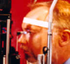
Ophthalmoscopy
During this examination, with the help of a special lens, we clinically
evaluate the optic nerve. In the picture, an optic nerve with glaucoma
of great cupping.

The above examinations may and must be performed not necessarily in a center that specializes in glaucoma, but in any ophthalmology clinic.
The study of visual fields is a very important examination which must always be performed if glaucoma is suspected. The patient must locate white light spots-targets on a white background.
 Pic.1: Automatic perimeter |
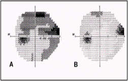 Pic.2: Visual fields with glaucoma |
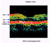
OCT 3
The OCT (Optical Coherence Tomography) is a new laser technology which
gives us a picture of tomographic transversal image of the retina layers.
With this technology we can study and measure the thickness of the nerve
fibers which are precosiously affected in glaucoma, as well as the parameters
of the optic nerve.
 |
 |
GDx
This technology allows us to evaluate and measure the thickness of the
nerve fiber layer and by consequence we make a first diagnosis since
it is the nerve fiber layer that first are affected by glaucoma.
 |
 |
Corneal pachymetry
The measurement of the thickness of the cornea is considered an important examination today since the thickness may influence the measurement of the pressure ie. the pressure may be overestimated in a thick cornea or underestimated in a thin cornea. Consequently we may get a wrong measurement of the pressure if we do not take into account the cornea’s thickness. The examination is performed with an ultrasonic pachymeter and only one contact with the cornea or with an optical pachymeter (ORBSCAN) during the cornea topography is enough.